
Understanding Professional Mole Evaluation and Dermoscopy
A detailed look at how dermatologists evaluate moles using dermoscopy, biopsy thresholds, and risk-based surveillance.
The Science Behind Mole Evaluation
Professional mole evaluation combines visual inspection, dermoscopy, and clinical judgment to distinguish benign moles from those that may be malignant. While the ABCDE rule gives patients a framework for self-examination, dermatologists apply a much more detailed analysis that includes pigment network patterns, streaks, blue-white veils, and atypical vascular structures visible only under magnification. The goal is to identify the small number of moles that warrant biopsy while avoiding unnecessary procedures on the vast majority that are harmless. A skilled evaluator can confidently dismiss many concerning-looking moles, sparing patients anxiety and scars.
How Dermoscopy Works
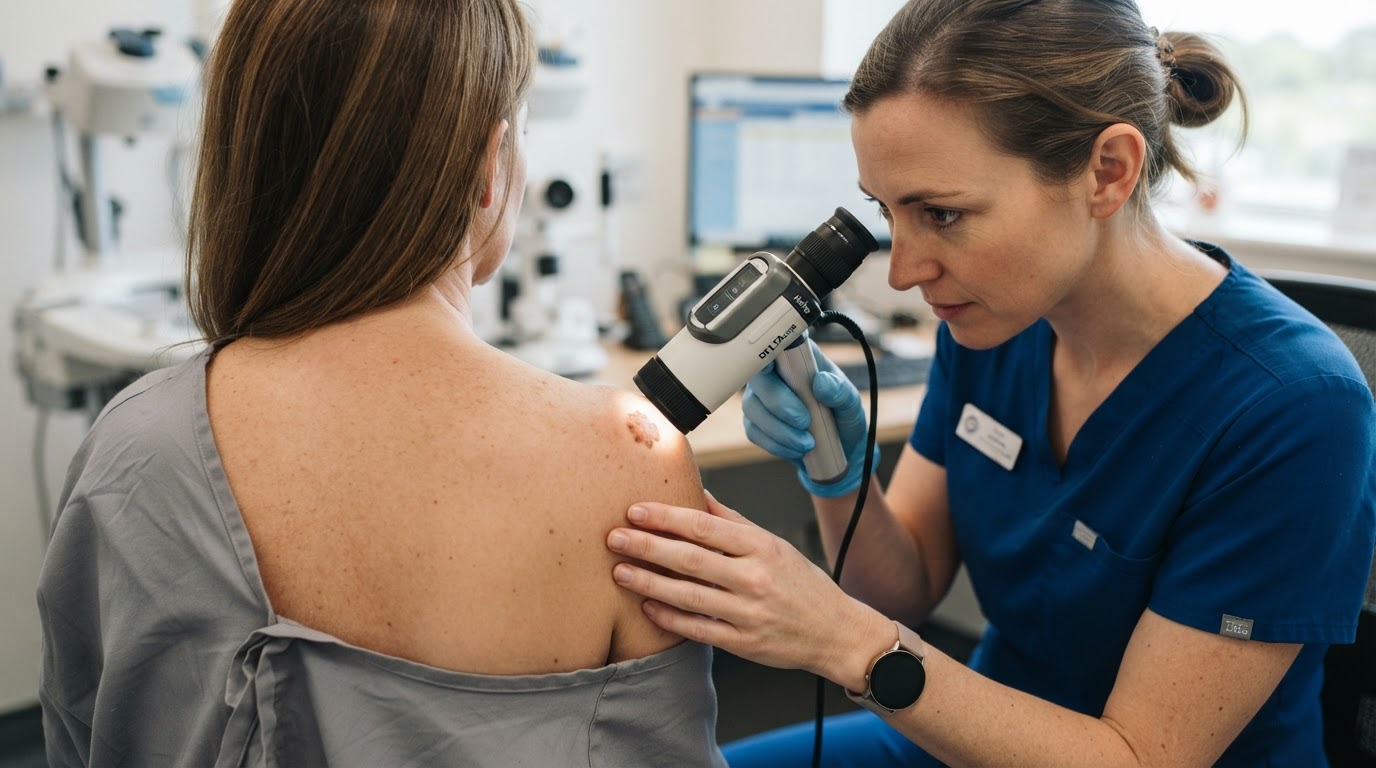
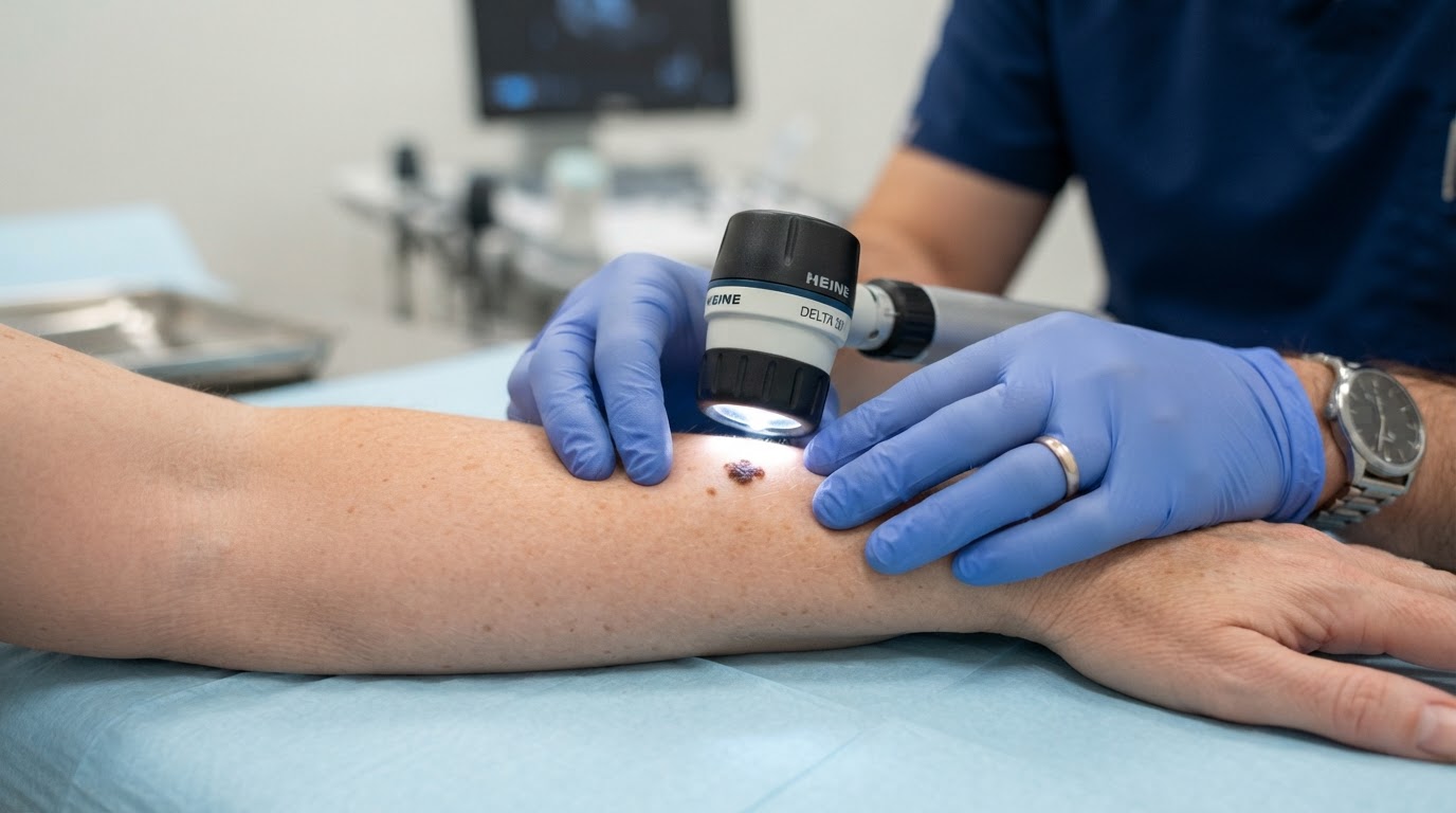
Dermoscopy uses a handheld device with polarized light and magnification to visualize structures beneath the skin surface that are invisible to the naked eye. The dermatologist presses the device against the skin or holds it just above, depending on the type of illumination, and examines the lesion's pigment network, dots, globules, streaks, and vessels. Specific patterns correlate with benign nevi, while others suggest melanoma. Polarized dermoscopy, in particular, reveals deeper structures without the need for immersion oil, making the examination quick and comfortable. Training in dermoscopy significantly improves a clinician's diagnostic accuracy compared with naked-eye inspection alone.
When a Biopsy Is Recommended
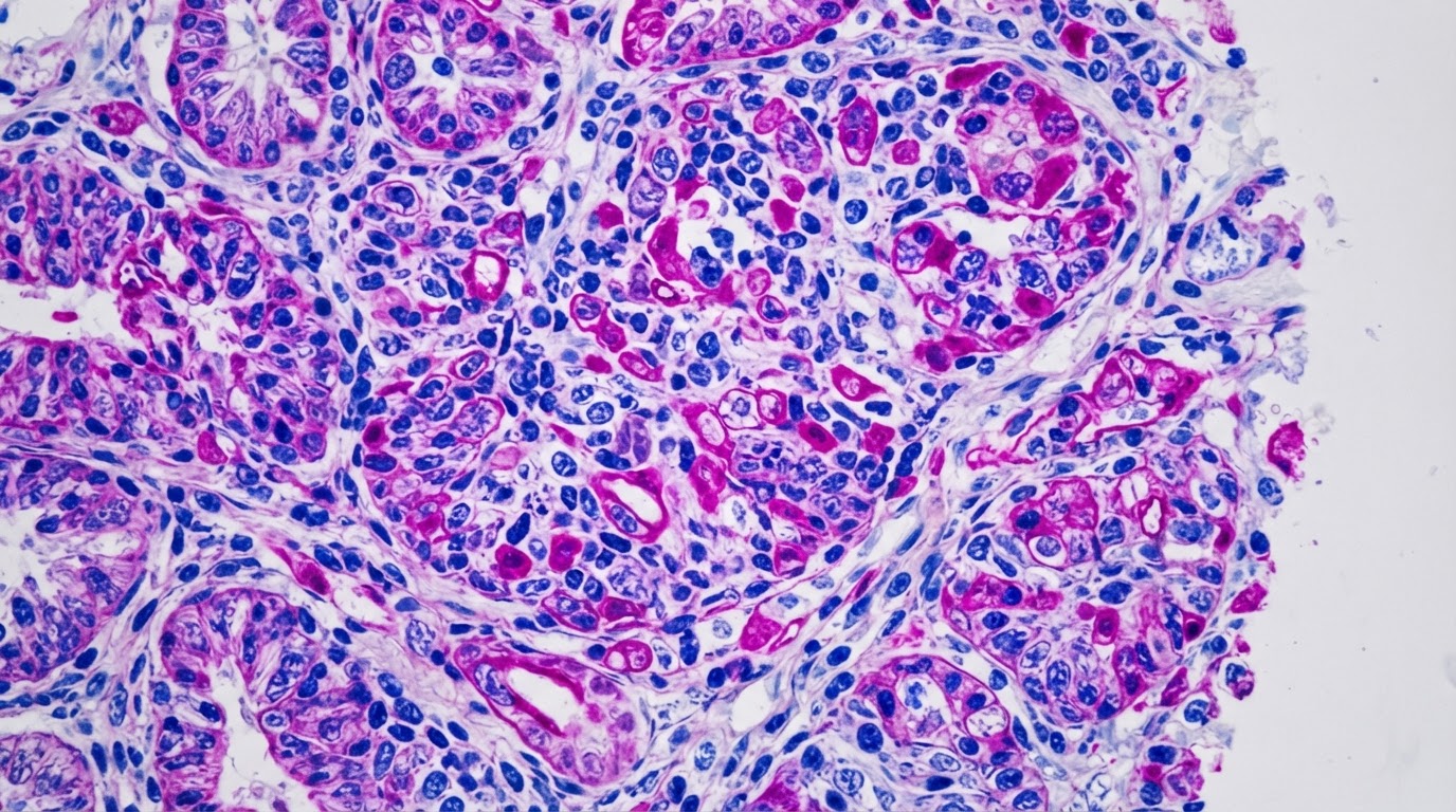
Not every unusual-looking mole requires a biopsy. The dermatologist weighs the dermoscopic findings against your personal risk factors, the mole's history of change, and its appearance relative to your other moles. A biopsy is typically recommended when a lesion shows chaotic pigment patterns, atypical vessel structures, or features that do not match the expected pattern for its location on the body. The most common biopsy techniques are the shave biopsy, which removes the raised portion of the lesion, and the punch biopsy, which removes a small full-thickness cylinder of skin. For lesions suspected to be melanoma, an excisional biopsy that removes the entire lesion is often preferred.
Risk-Based Surveillance After Evaluation
Following a mole evaluation, the dermatologist will recommend a surveillance plan tailored to your risk profile. Patients with a personal history of melanoma, numerous atypical moles, or a strong family history typically require examinations every three to six months. Patients with average risk may be advised to return annually. If total body photography or digital mole mapping is available, the dermatologist may use it to track subtle changes over time, which is particularly valuable for patients with many moles. Understanding your surveillance plan helps you stay engaged in your own preventive care.
Partnering with Your Dermatologist
The most effective mole evaluation happens when you and your dermatologist work together. Arrive with a list of any moles that have changed, note when the change began, and be honest about your sun exposure history and any prior skin procedures. Between visits, perform monthly self-examinations and report any new or evolving spots promptly. Professional mole evaluation is most powerful when it is part of an ongoing relationship rather than a one-time event, and consistent surveillance catches changes at the earliest, most treatable stage.
Tracking Moles Between Visits
Between professional evaluations, your own observations are a crucial part of effective surveillance. Set a recurring monthly reminder to perform a self-examination in a well-lit room, using a full-length mirror alongside a hand mirror to inspect the back, scalp, and other hard-to-see areas. For patients with numerous moles, total body photography can be invaluable: taking standardized photographs of the entire skin surface every six to twelve months creates an objective baseline that makes even subtle changes obvious at the next comparison. Some dermatology practices offer digital mole mapping, which tracks individual lesions over time and flags those that have changed dimension or pigment pattern. If you notice a mole that meets any of the ABCDE criteria, or that simply looks different from the others on your body, do not wait for your next scheduled appointment. Contact your dermatologist promptly, because early evaluation of a changing mole is far more valuable than watchful waiting. Combining professional dermoscopy with disciplined self-tracking gives you and your dermatologist the information needed to act quickly when it matters most.
This article is educational and is not a substitute for professional medical advice, diagnosis, or treatment. Always seek the guidance of a qualified dermatologist or other licensed health provider with questions about a medical condition.
Looking for a specialist in this area? Browse Skin Biopsies & Dermatopathology