
Squamous Cell Carcinoma Treatment Options Explained
From cryotherapy to Mohs surgery and radiation, a practical guide to treating squamous cell carcinoma of the skin.
What Squamous Cell Carcinoma Is
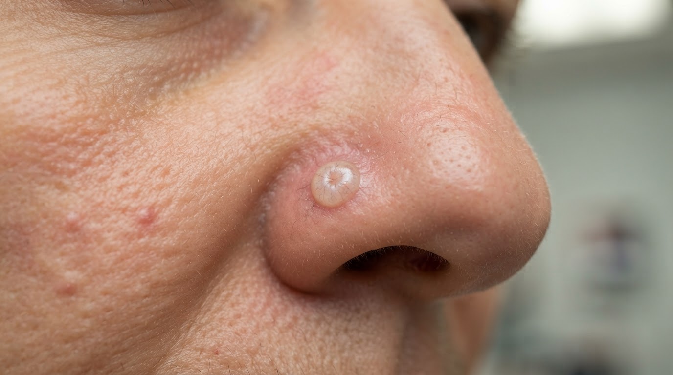
Squamous cell carcinoma is the second most common form of skin cancer, arising from the squamous cells that make up the upper layer of the epidermis. It most often appears on sun-exposed skin such as the face, ears, scalp, hands, and arms, frequently developing from a precancerous actinic keratosis. Unlike basal cell carcinoma, squamous cell carcinoma has a small but real potential to spread to lymph nodes and other organs, particularly when it occurs on the lip, ear, or in immunosuppressed patients. Prompt, appropriate treatment is important both to cure the local tumor and to prevent metastasis.
Treating Precursor Actinic Keratoses
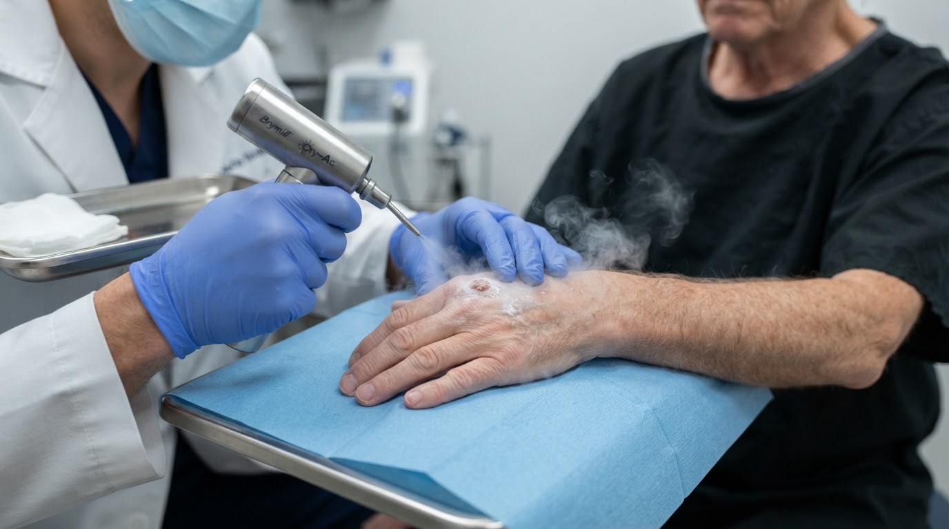
Many squamous cell carcinomas begin as actinic keratoses, rough scaly spots that develop on chronically sun-damaged skin. Treating actinic keratoses prevents progression to squamous cell carcinoma. Options include cryotherapy with liquid nitrogen, topical creams such as 5-fluorouracil and imiquimod, photodynamic therapy, and chemical peels. Field therapies that treat a broad area of sun damage are particularly useful for patients with numerous actinic keratoses. Regular treatment of precancerous lesions is one of the most effective ways to reduce the risk of developing invasive squamous cell carcinoma.
Surgical Excision and ED&C
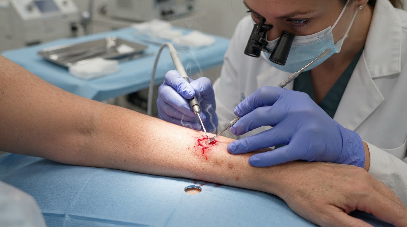
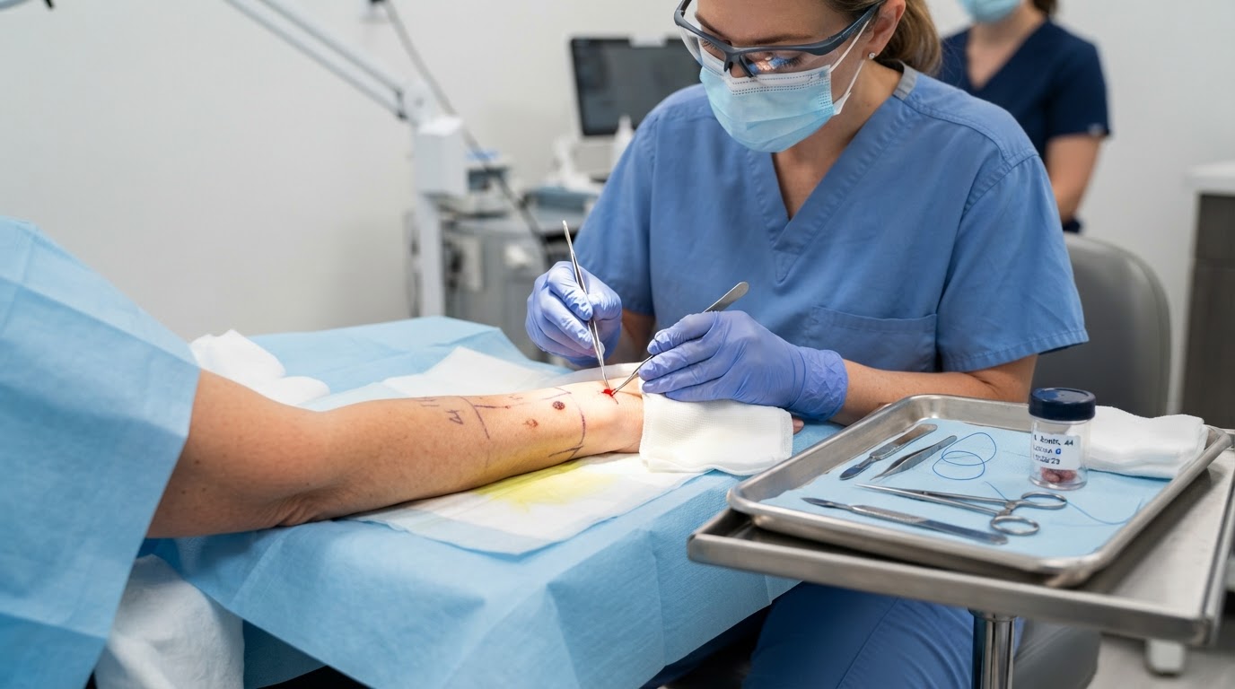
For invasive squamous cell carcinoma, surgical excision is the most common treatment. The dermatologist removes the tumor with a margin of healthy skin, and the specimen is examined pathologically to confirm clear margins. Electrodessication and curettage can be used for small, low-risk lesions on the trunk or limbs, though it is less appropriate for higher-risk areas such as the lip or ear. Both procedures are performed in the office under local anesthesia. The choice depends on tumor size, location, depth, and histologic features such as perineural involvement, which can indicate a more aggressive course.
Mohs Surgery for Higher-Risk Lesions
Mohs micrographic surgery is recommended for squamous cell carcinomas in areas where tissue conservation is important, such as the face, lips, ears, and hands, and for tumors that are large, recurrent, or have aggressive histologic features. By examining the entire margin microscopically, Mohs achieves the highest cure rate while sparing healthy tissue, which is especially valuable on the face where function and appearance matter. For squamous cell carcinoma with perineural invasion or other high-risk features, Mohs may be combined with radiation therapy and a multidisciplinary evaluation to ensure complete management.
Radiation and Systemic Options
Radiation therapy is an option for patients who cannot undergo surgery, for tumors in difficult locations, or as an adjunct after excision of high-risk lesions. For squamous cell carcinoma that has spread to lymph nodes or beyond, systemic therapies including immunotherapy and chemotherapy may be coordinated with a medical oncologist. Immunosuppressed patients, such as organ transplant recipients, require particularly close surveillance because their risk of squamous cell carcinoma is dramatically elevated. The right treatment plan depends on the tumor's risk features and your overall health, and a dermatologist experienced in skin cancer care will guide you through the options to achieve the best possible outcome.
Follow-Up and Prevention After Treatment
Recovery from squamous cell carcinoma treatment extends well beyond the day of the procedure. Most surgical sites heal within two to four weeks, and your dermatologist will give you detailed wound-care instructions covering cleaning, ointment, and signs of infection to watch for such as increasing redness, warmth, or pain. Sun protection is essential both to support healing and to reduce the risk of future skin cancers, so keep the treated area covered and apply a broad-spectrum sunscreen daily once the skin has closed. Because anyone who has had one squamous cell carcinoma faces a higher risk of developing another, ongoing surveillance is a central part of long-term care. Your dermatologist will recommend a follow-up schedule, often a full-body skin examination every six to twelve months, and more frequent visits for tumors that had high-risk features or for patients who are immunosuppressed. Between professional visits, perform a monthly self-examination and report any new scaly patches, firm nodules, or non-healing sores promptly. For patients with numerous actinic keratoses, regular field treatment of precancerous lesions helps prevent new squamous cell carcinomas from forming. Combining diligent sun protection, consistent professional surveillance, prompt attention to new lesions, and proactive treatment of precancers gives you the strongest protection against recurrence and keeps your skin health on track for the long term.
This article is educational and is not a substitute for professional medical advice, diagnosis, or treatment. Always seek the guidance of a qualified dermatologist or other licensed health provider with questions about a medical condition.
Looking for a specialist in this area? Browse Basal & Squamous Cell Carcinoma Care