
Basal Cell Carcinoma Treatment Options Explained
A clear breakdown of how dermatologists treat basal cell carcinoma, from topical creams to Mohs surgery and targeted therapy.
Understanding Basal Cell Carcinoma
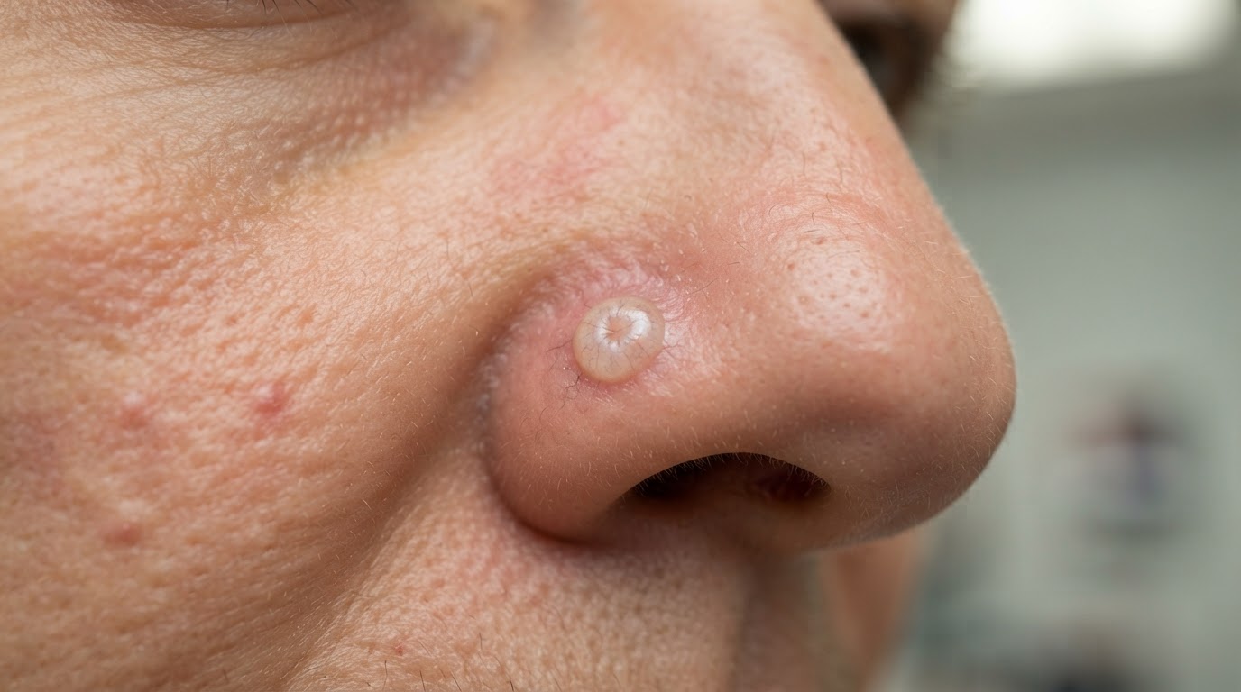
Basal cell carcinoma is the most common form of skin cancer and the most common cancer overall in the United States. It arises from the basal cells at the base of the epidermis, usually on skin that has accumulated years of sun exposure such as the face, ears, neck, scalp, and arms. Basal cell carcinoma grows slowly and rarely spreads beyond the skin, but it can cause significant local damage if left untreated, invading cartilage, bone, and other nearby structures. Because it is so common and so treatable when caught early, understanding your treatment options is an important part of managing a diagnosis.
Topical Treatments for Superficial Lesions
For very superficial basal cell carcinomas, particularly those on the trunk or limbs where surgery would leave a larger scar, topical treatments can be effective. Prescription creams such as imiquimod and 5-fluorouracil trigger an immune or inflammatory response that destroys the cancer cells over several weeks of application. These treatments require diligence and follow-up to confirm clearance, and they are not appropriate for deeper or more aggressive tumors. Your dermatologist will consider the tumor's thickness, location, and histology before recommending a topical approach over surgery.
Surgical Excision and ED&C
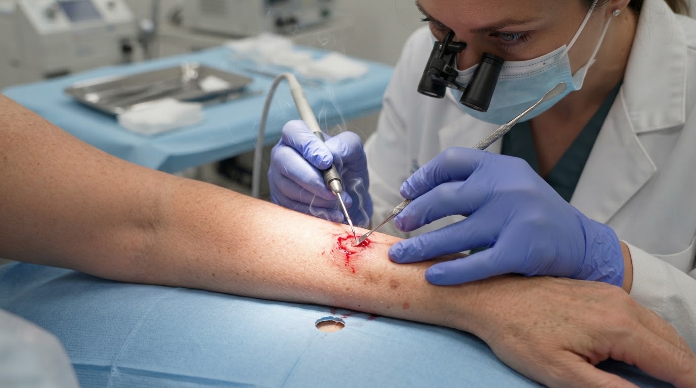
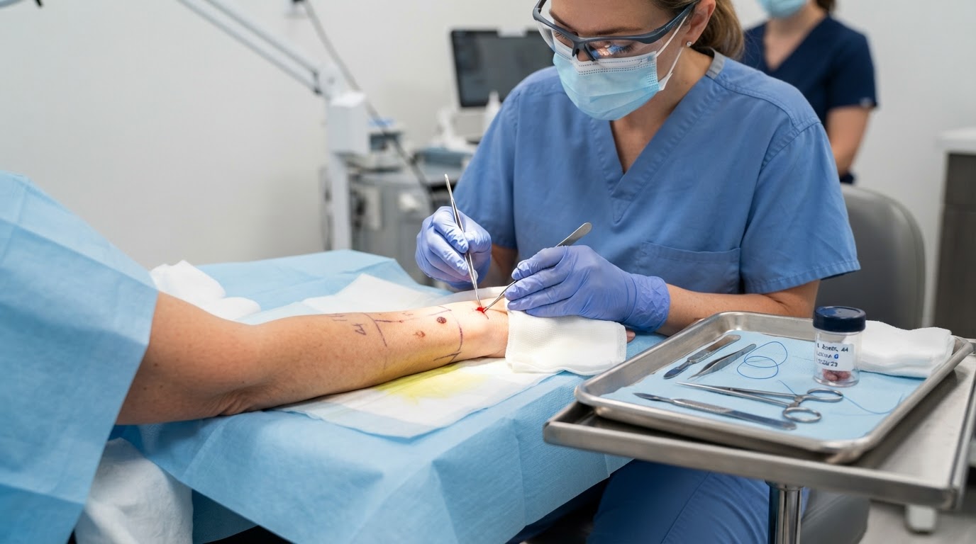
Surgical excision is the most common treatment for basal cell carcinoma. The dermatologist removes the visible tumor along with a margin of healthy skin, and the specimen is sent to a pathology lab to confirm that the margins are clear. Electrodessication and curettage, or ED&C, is a quick scrape-and-burn technique suitable for small, superficial lesions in low-risk areas. Both procedures are performed in the office under local anesthetic and have high cure rates for appropriately selected tumors. The choice depends on tumor location, size, and the cosmetic outcome you prefer.
Mohs Micrographic Surgery
Mohs micrographic surgery is the preferred treatment for basal cell carcinomas on the face and other areas where preserving healthy tissue is critical. By examining one hundred percent of the surgical margins under a microscope while the patient waits, the Mohs surgeon confirms complete removal and achieves cure rates up to ninety-nine percent for new tumors. Mohs is especially valuable for recurrent basal cell carcinomas, large tumors, and aggressive histologic subtypes such as infiltrating or morpheaform basal cell carcinoma. Although the procedure takes longer than standard excision, the precision and tissue sparing are unmatched.
Advanced and Targeted Therapies
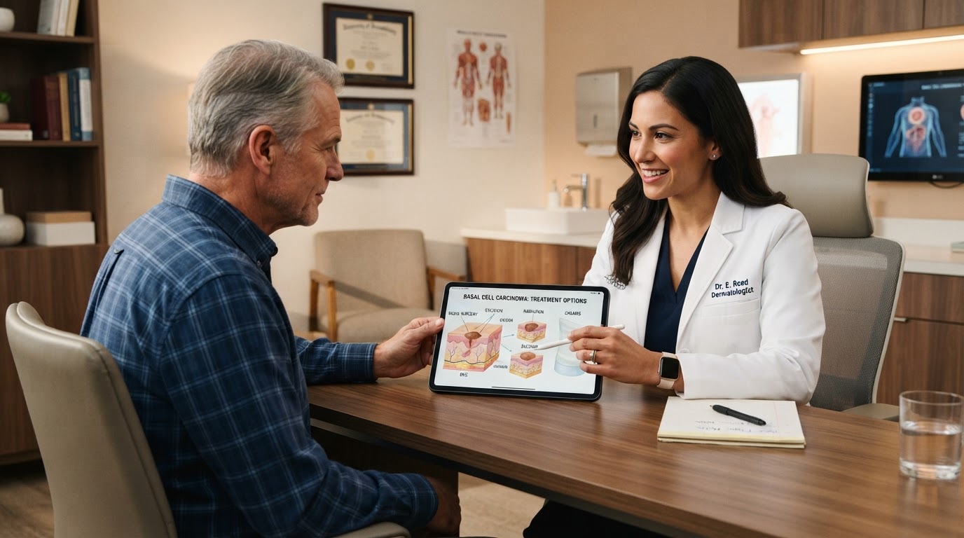
For the small number of basal cell carcinomas that are advanced or inoperable, targeted oral therapies known as hedgehog pathway inhibitors can shrink tumors that cannot be removed surgically. Radiation therapy is another option for patients who cannot undergo surgery due to age or medical condition. These advanced approaches are coordinated with specialists and are reserved for cases where standard surgical treatment is not feasible. Most basal cell carcinomas, however, are cured with straightforward surgical or topical treatment, and the key to a good outcome is early detection and a treatment plan tailored to the specific characteristics of your tumor.
Recovery and Long-Term Surveillance
After basal cell carcinoma treatment, attention turns to healing and to preventing future skin cancers. Most surgical sites heal within two to four weeks, and your dermatologist will provide specific wound-care instructions, including how to keep the area clean, when to apply ointment, and how to recognize signs of infection such as increasing redness, swelling, or pain. Sun protection is essential both for healing and for prevention, so keep the treated area covered and apply a broad-spectrum sunscreen daily once the skin has closed. Because a person who has had one basal cell carcinoma has a substantially higher risk of developing another, ongoing surveillance is a critical part of long-term care. Your dermatologist will recommend a follow-up schedule, often a full-body skin examination every six to twelve months, to catch any new primary tumors at the earliest stage. Between visits, perform monthly self-examinations and report any new pearly bumps, non-healing sores, or changing spots promptly. Combining diligent sun protection, consistent professional surveillance, and prompt attention to new lesions gives you the best chance of staying skin-cancer-free after treatment, and it turns a single diagnosis into a manageable, long-term preventive routine.
This article is educational and is not a substitute for professional medical advice, diagnosis, or treatment. Always seek the guidance of a qualified dermatologist or other licensed health provider with questions about a medical condition.
Looking for a specialist in this area? Browse Basal & Squamous Cell Carcinoma Care