
What Happens During a Professional Skin Cancer Screening
A step-by-step walkthrough of a clinical skin cancer screening, from intake to dermoscopy, so you know exactly what to expect.
The Purpose of a Skin Cancer Screening
A professional skin cancer screening is a head-to-toe visual examination performed by a trained clinician to identify suspicious lesions before they become dangerous. Unlike a self-exam in your bathroom mirror, a clinical screening uses bright lighting, magnification, and dermoscopy to reveal pigment patterns and vascular structures that are invisible to the naked eye. The goal is not only to find cancer that is already present but to establish a baseline against which future changes can be measured. For adults with fair skin, a history of sunburns, or a family history of melanoma, regular screening is one of the most effective preventive measures available.
Before the Examination Begins
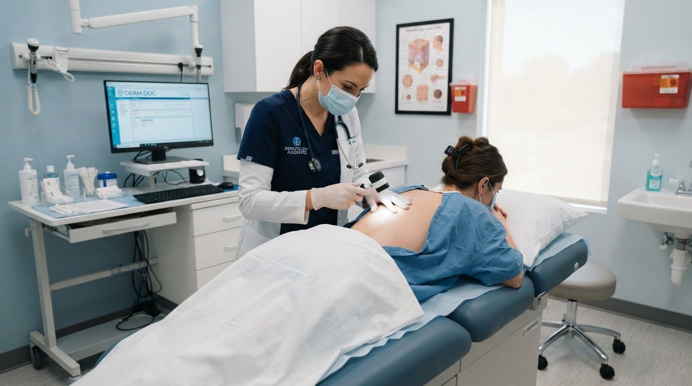
When you arrive, a medical assistant will record your height, weight, and relevant medical history, including any personal or family history of skin cancer, immunosuppressive medications, and prior biopsies. You will be asked to remove your clothing down to your undergarments and put on a medical gown that allows access to the entire skin surface. It is important to remove nail polish and avoid makeup on facial lesions so nothing obscures the doctor's view. The clinician will ask whether you have noticed any particular spots of concern, so arrive with a brief list ready.
The Full-Body Examination Itself
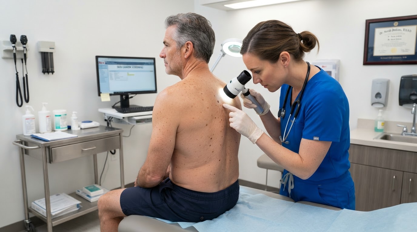
The dermatologist will systematically inspect your skin from your scalp to the soles of your feet, including areas you may never check yourself such as the scalp between the hairs, behind the ears, between the toes, and the buttocks. The examination typically takes only a few minutes for a patient with no prior history, though it may be longer for someone with numerous moles. The doctor is looking for the ABCDE warning signs of melanoma, pearly bumps that suggest basal cell carcinoma, and scaly patches that may indicate squamous cell carcinoma or actinic keratosis.
Dermoscopy: Looking Beneath the Surface
When the dermatologist identifies a spot that looks unusual, they will use a dermatoscope, a handheld magnifying device with polarized light, to examine the lesion in detail. Dermoscopy reveals structures such as pigment networks, streaks, blue-white veils, and atypical vessels that help distinguish benign moles from malignant ones. This non-invasive technique dramatically improves diagnostic accuracy compared with naked-eye inspection alone and often prevents unnecessary biopsies. A skilled dermatologist can confidently dismiss many concerning-looking spots with dermoscopy, sparing you a procedure.
What Comes Next
If the screening reveals a lesion that warrants further evaluation, the dermatologist will recommend a biopsy, usually performed the same day or scheduled within a few days. If nothing suspicious is found, you will be advised on how often to return for follow-up based on your personal risk profile. You will also receive guidance on sun protection and self-examination between visits. Understanding the process in advance removes much of the anxiety from a screening and helps you arrive prepared, which improves the quality and value of the visit.
Building a Long-Term Screening Habit
A single screening is valuable, but the real protection comes from making skin cancer screening a recurring part of your preventive health routine. Adults at average risk often benefit from an annual screening, while those with a personal history of skin cancer, numerous atypical moles, significant sun exposure, or a family history of melanoma may need to return every three to six months. Ask your dermatologist for a written surveillance schedule at the end of each visit so you can plan ahead rather than waiting until a concern arises. Between professional visits, perform a monthly self-examination using a full-length mirror and a hand mirror for hard-to-see areas such as the back of the scalp and the backs of the legs. Document any new or changing spots with a photograph and the date, which gives your dermatologist objective evidence of evolution at your next appointment. Combining consistent professional screening with diligent self-examination is the most reliable way to catch skin cancer at its earliest and most treatable stage.
This article is educational and is not a substitute for professional medical advice, diagnosis, or treatment. Always seek the guidance of a qualified dermatologist or other licensed health provider with questions about a medical condition.
Looking for a specialist in this area? Browse Full-Body Skin Examinations & Screening